

BY SUSAN FOX, DO
Venous stasis ulcers are the most common form of leg ulcerations. As much as 80% of lower extremity ulcers are due to venous disease. Venous disease exists in 25% of the U.S. population as a whole. Vein disease affects 70% of all women and 40% of all men. More than one million Americans have venous stasis ulcers, and more than 100,000 people are totally disabled as a result. One-quarter of patients have their first ulcers by 40 years of age and three-quarters by 60 years of age.
The average cost to treat a venous ulcer is approximately $10,000, resulting in a huge burden on the U.S. health care system — $2.5 billion to $3 billion each year. Between three and four million workdays are lost annually. As much as 70% of venous ulcers recur within the first six months to a year of healing. It is important to diagnose venous insufficiency early in its course and treat it appropriately before the patient develops an ulcer.
Those at Risk
Several epidemiological studies have been performed to determine who develops venous insufficiency. Older age, heredity, obesity, a history of phlebitis or leg trauma, surgery, or crush injuries are major risk factors. Debate exists over whether professions that require people to work on their feet for the majority of the day (i.e., nurses, hairdressers, teachers) help contribute to venous insufficiency.
The majority of ulcers are due to chronic venous insufficiency. When a person is standing upright, blood returning to the heart from the lower extremities must travel against gravity. As one walks, calf muscles contract, compressing the deep veins and propelling blood upward. Normally, semilunar valves in the veins prevent the backflow of blood. However, valvular failure and muscle weakness can lead to the backflow of blood. This retrograde flow of blood leads to pooling of blood in the veins and venous hypertension.
These elevated venous pressures communicate to the superficial veins and cause the veins to stretch, creating superficial varices. The elevated pressures cause even the thin-walled capillaries (small veins) to stretch, leaving gaps between the cell walls. As the single-layer-cell walled capillaries stretch, water, proteins (causing fibrin cuffs), and red blood cells leak through these gaps, causing leg edema, tissue changes, and discoloration around the ankles.
Progression
Venous disease is broken down into three stages. Stage one is edema in the anterior shin and ankles with starburst spider vein formations around the medial ankle. Stage two is discoloration from hemosiderin (an iron pigment in the blood) staining with swelling of the lower extremity. Gradually, skin hardening, called lipodermatosclerosis, occurs, often containing thin white atrophic scars and small painful foci primarily on the medial ankle or posterior foot. The exact step leading from venous hypertension to ulceration is unknown. In stage three, an ulcer develops primarily in the medial ankle, but as much as 20% may occur in other areas.
Venous ulcers are often located around the medial malleolus in an area referred to as the gaiter area. Skin around the ulcer tends to be swollen and pigmented. Venous ulcers are often larger than arterial ulcers. Patients should undergo baseline arterial testing with and without exercise to rule out concomitant arterial disease. Also, a venous color duplex ultrasound is needed. A venous insufficiency study is important to determine the exact location of valvular incompetence and any possible arteriovenous shunts or thrombosis. Invasive phlebography is rarely if ever used now to diagnose venous disease.
The traditional, conservative modality used to treat venous ulcerations is compression therapy (Unna boots, Dyna-Flex™, Profor, multilayer compression bandages, compression hose, or soft casting). As long as the arterial supply to the lower extremity is intact, multilayer compression bandages or medical-strength compression hose are applied to the lower extremity to decrease lower extremity edema.
ELVeS™ is one of the newer options for treating venous leg ulcers. This endovenous laser treatment is minimally invasive and results in less postoperative pain, fewer wound infections, fewer scars, fewer missed varices, fewer recurrences, fewer nerve injuries, and fewer days off work than vein-stripping surgery.
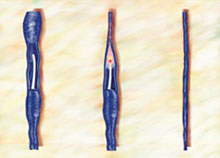
| Varicose vein before treatment; ELVeS laser and catheter inserted into vein | Laser beam heats interior of varicose vein; vein scars down and collapses as catheter is withdrawn | ELVeS catheter is removed; vein remains closed and shrinks over time |
Leg elevation, moisturizing the lower extremity, and good foot care also play key roles. Having patients sleep with their legs elevated 4 to 6 inches reduces the majority of edema by morning. Before getting out of bed, the person should immediately apply medical grade compression hose. The compression hose keep the edema out of the extremity and allow nutrients in the body to heal the damaged tissue. Calf-pumping exercises, in addition to the compression therapy, enhance venous return and facilitate ulcer healing. Even after the ulcer is healed, patients need to continue to wear compression hose on their legs. Patients who are compliant with wearing compression hose have increased ulcer healing rates and decreased recurrence rates.
Beyond Vein Stripping
Because 70% of venous ulcers recur, patients with symptoms of venous insufficiency (leg pain, swelling, discoloration, malleolar flare, and tissue damage) with or without leg ulceration should be evaluated for one of the new treatment options to treat their venous disease definitively. This early treatment can help prevent development of venous ulcers or their recurrence.
The conventional treatment for chronic venous disease was vein stripping. Now, newer treatment options are available, such as endovenous laser and radiofrequency ablation of the veins. These newer, minimally invasive treatment modalities result in less postoperative pain, fewer wound infections, fewer scars, fewer missed varices, fewer recurrences, fewer nerve injuries, and fewer days off work than vein-stripping surgery. Recovery takes just a few days. The radiofrequency ablation has a success rate of 90% at two years, and the endovenous laser has success rates ranging from 96% to 100% at two years. The procedures close off the superficial incompetent vein(s) and are usually performed in the office under local anesthetic.
Whether the venous disease is due to superficial or deep venous insufficiency, new treatment options are available. Studies have shown that intensive treatment of superficial venous disease, both varicose and spider veins, can improve deep venous flow. In one study, more than 80% of deep venous blood flow improved (resolving venous insufficiency and shrinking vein size) after aggressive treatment of the superficial veins. This improvement in blood flow assists in the healing of venous stasis ulcers and decreases their recurrence. Aggressively treating venous disease helps prevent venous ulcerations that are a huge health care burden.